Training, long-term and short-term anabolic steroid use ---effect on muscle fibers
Recently I posted about a well-known fact, we now call “muscle memory”. Underneath this article you can find the links to these blog posts.
A high-level power-lifter from Sweden that happens to be a scientific researcher was fascinated by this process and wanted to know what long-term (10 years and longer) short-term (cycle of 20 weeks) did to muscles compared to natural power-lifters.
He compared his research with other similar studies. He also wanted to know what the difference could be of training with or without anabolic steroids long term or short term to an isometric contraction such as the dead-lift compared to a maximal explosive exercise such as the squat.
First an overview of the muscle fibers involved.
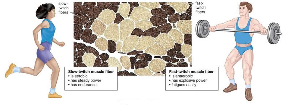
There are three primary muscle fiber types in humans -- Type I, Type IIA, and Type IIB. Type I are referred to as "slow twitch oxidative", Type IIA are "fast twitch oxidative" and Type IIB are "fast twitch glycolytic" As their names suggest, each type has very different functional characteristics. Type one fibers are characterized by low force/power/speed production and high endurance, Type IIB by high force/power/speed production and low endurance, while Type IIA fall in between. These characteristics are a result, primarily, of the fiber's Myosin Heavy Chain (MHC) composition, with Mysosin heavy chain isoforms I, IIa and IIx corresponding with muscle fiber types I, IIA, and IIB.
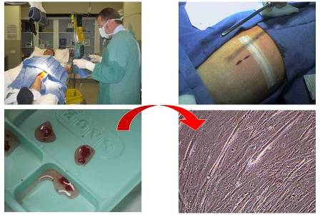
Individual muscles are made up of individual muscle fibers and these fibers are further organized into motor units grouped within each muscle. A motor unit is simply a bundle or grouping of muscle fibers. When you want to move the brain nearly instantaneously sends a signal or impulse through the spinal cord that reaches the motor unit. The impulse then tells that particular motor unit to contract it's fibers. When a motor unit fires all the muscle cells in that particular motor unit then contract with 100% intensity. So, a muscle cell either contracts 100% or not at all. A motor unit is either recruited 100% or not at all. Therefore, there is no such thing as a partially firing motor unit or a partially contracted muscle fiber.
The body recruits the lower threshold motor units first (slow-twitch), followed by the higher threshold motor units (fast-twitch) and continues to recruit and fire motor units until you’ve applied enough force to do whatever it is you’re trying to do regarding movement. When you are lifting something extremely heavy or applying a lot of force your body will contract practically all the available motor units for that particular muscle.
When engaging in high intensity or high force activities you get lots of motor unit activation and thus a lot of force. So how does this relate to the fiber in the available motor units? Well type I muscle motor units contract less forcefully and a little slower than type II fast twitch motor units and they reach peak power slower. They are also highly resistant to fatigue so they have good endurance. This is why you can sit and eat all day or play Playstation all day and never get tired!
The type II motor units are divided into type IIA and type IIB. Both of these sub-groups are capable of greater levels of absolute force than type I and also fatigue a lot quicker. Type IIA and IIB are capable of roughly the same amount of peak force, but the IIA fibers take longer to reach their peak power in comparison to type IIB. Type IIA fibers reach peak power in about 50 milliseconds whereas type IIB reaches peak power in about 25 milliseconds. Because of their greater contraction speeds, the total peak power by IIB can be up to 5 times higher than the IIA's.
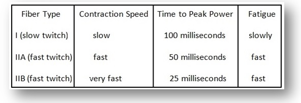
Now, when we realize that sports movements usually occur in around 200 milliseconds or less, if you look at the time to peak power of the individual muscle fibers, it should then become obvious that each type (I,IIA,IIB) has enough time to reach peak power production. So, why the superiority in having more fast twitch II B fibers? Well, two things. Since they contract quicker, if you have an advantage for the first tenth (arbitrary) of the movement, it can result in superior performance. Since their total peak power is greater this could also give one an advantage when producing force under high velocity conditions.
From the view of being successful in sports, examining the individual data for the subjects revealed that each individual athlete actually showed a personal muscle profile. A person with higher proportion of type IIB fibers would, from a theoretical point of view, have a greater chance of becoming a successful power lifter.
Power-lifters on long-term anabolic steroid supplementation (PAS)
Power-lifters never taking anabolic steroids (P)
Vastus lateralis (VL)
Trapezius muscle (T)
The morphological appearance of the vastus lateralis (VL) muscle from high-level power-lifters on long-term anabolic steroid supplementation (PAS) and power-lifters never taking anabolic steroids (P) was compared. The effects of long- and short-term supplementation were compared. Enzyme-immunohistochemical investigations were performed to assess muscle fiber type composition, fiber area, number of myonuclei per fiber, internal myonuclei, myonuclear domains and proportion of satellite cells. The PAS group had larger type I, IIA, IIAB and IIC fiber areas . The number of myonuclei/fiber and the proportion of central nuclei were significantly higher in the PAS group (pThe initial effects from AS appear to be maintained for several years.
//juicedmuscle.com/jmblog/content/muscle-memory-fact-or-fiction
//www.juicedmuscle.com/jmblog/content/steroids-probably-result-lasting-benefit-its-users
Investigating the mechanisms of long-term supplementation of AS on human trapezius muscle, Kadi ( 2000) showed that the enlargement of muscle fibers is accompanied by an increase in the myonuclear number and satellite cell content. In addition the PAS group have a significant higher number of cells expressing fetal myosin heavy chain (MyHC) than power lifters not using anabolic steroids, indicating ongoing new fiber generation.
The alterations in the number of myonuclei and satellite cells in response to AS were later confirmed in a study on the short term effects (20 weeks) of combined treatment with gonadotropin-releasing hormone (GnRH) agonist and testosterone on the vastus lateralis muscle (Sinha-Hikim et al. 2002, 2003). However, the magnitude of muscle fiber hypertrophy was higher in trapezius from athletes with long-term AS supplementation than in muscles from athletes after 20 week of AS supplementation.
Another difference was the increased number of centrally located myonuclei in the trapezius muscle (Kadi 2000). These findings might reflect discrepancies in either the response to AS or differences in contraction pattern during exercise between the trapezius and the vastus lateralis muscles.
The vastus lateralis is of importance preferentially in the squat event, when the lifter from an upright position and with the barbell resting across the back of the shoulders, sits or ‘‘squats’’ down by doing a flexion in the knee and hip-joints to a required depth. The lifter then attempts to stand up again, returning to the original position. Such a lift takes approximately 2–5 s and requires a maximal explosive force.

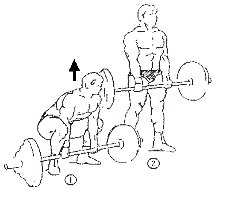
In a dead lift the lifter takes the barbell from the floor to an upright standing position until validated by the referee. In this lift the trapezius muscle performs an isometric contraction for a total time of approximately 8–10 s. These differences in utilization between vastus lateralis and trapezius might be reflected at the muscle fiber level.
The aim of the present investigation was to study the long-term effects of AS on fiber type composition, fiber area, number of myonuclei, internal nuclei, nuclear do-mains and the number of satellite cells in human vastus lateralis. We also compared the long-term effects of AS on vastus lateralis to the long-term effects of AS on the trapezius muscle, previously reported by Kadi et al. ( 1999,2000) and to the short-term effects (20 weeks) of AS on the vastus lateralis muscle (Sinha-Hikim et al. 2003).
Fiber types
A mosaic pattern of fiber types in cross-sectioned biopsies was observed in most subjects. The mean values for all fiber types in both groups were very similar but the individual variation was large . Type I and type IIA fibers were the most frequent fiber types in most subjects but their frequency varied from 28 to 62% for type I fibers and from 4 to 69% for type IIA fibers. The proportions of type IIAB and type IIB fibers varied even more. Type IIAB was found in proportions from 0 to 33% and type IIB from 0 to 50%. Interestingly, type IIB fibers occurred preferentially in two subjects in the P group. Fiber type grouping occurred in several of the biopsies from subjects in the PAS group, but also to some extent in biopsies from subjects in the P group.
No statistically significantly differences were observed in the proportions of fiber types between the PAS and the P groups.
Fiber area
Type IIA and type IIAB fibers had the largest mean fiber area in both groups, but the PAS group had 44% larger type II fibers than the P group (p
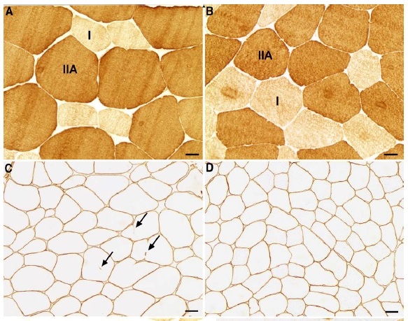
Immunostained crossections of one PAS subject (A and C) and two P subjects (B and D).
Discussion
To our knowledge, this is the first comparative morphological study of the human vastus lateralis muscle in two groups of high-level power-lifter athletes. One group admitted supplementation with testosterone, anabolic steroids and other banned substances for nearly ten years and the other group affirmed never taken banned substances. As we previously have studied the trapezius muscle from the same individuals (Kadi et al. 1999b) it allows us now to compare the AS influence on two muscles used in different ways in power lifting.
The vastus lateralis muscle demonstrated larger muscle fiber areas than normal in both groups (Staron et al. 2000). However the PAS subjects differed from the P subjects in having significantly larger fiber areas, more myonuclei per fiber and more internal myonuclei. No differences were seen with regard to fiber type proportions and frequency of satellite cells. All subjects in the PAS group and seven in the P group had small fibers expressing fetal myosin but there was no significant difference between the groups.
The PAS group had 61% larger type I fiber area and 44% larger type II fiber area than the P group. In the trapezius, the areas were 58 and 33% larger in type I and type II fibers, respectively (Kadi et al. 1999b). In fact, AS supplementation, even without strength training has been reported to induce hypertrophy in human skeletal muscles (Bhasin et al. 1996; Sinha-Hikim et al. 2002). In the study by Sinha-Hikim et al. ( 2002) the muscle fiber hypertrophy (fiber area) after 20 weeks of AS supple-mentation (600 mg/week) was 49% in type I fibers and 36% in type II fibers compared to baseline. The dramatic hypertrophic effect on muscle fibers in subjects supplementing with AS is in accordance with the current conception on the effects of testosterone and anabolic steroids, for review see (Herbst and Bhasin 2004). Maximal force of a muscle is related to the muscle fiber area, the total muscle area and the fiber types (Bruce et al. 1997; Bamman et al. 2000).
In the present study we observed that there was a highly significant positive correlation between fiber area and total number of myonuclei in the vastus lateralis muscle. The PAS subjects, who had the largest fibers, also had the largest numbers of myonuclei, both subsarclemmal and centrally located. The same correlation was observed in our study on the trapezius muscle (Kadi et al. 1999b). In the study from Sinha-Hikim et al. ( 2002) the number of myonuclei was significantly increased and was correlated to fiber area. Altogether, these results support the idea that the number of myonuclei plays a mechanistic role in muscle fiber hypertrophy (Edgerton and Roy 1991; Allen et al. 1999; Kadi 2000).
It is well known that each nucleus supports a certain volume of the cytoplasm with mRNA for turnover of proteins. This volume is often referred to as a nuclear domain (Cheek 1985). If a myonucleus can expand its nuclear domain by increased synthesis of mRNA or by more efficient transport of mRNA has been discussed (Sinha-Hikim et al. 2003). Recently Kadi et al. ( 2004) reported that satellite cells are plastic in response to resistance training and that moderate changes in skeletal muscle fiber area can be achieved without addition of new myonuclei. However, our data suggest that addition of myonuclei is a prerequisite for more substantial muscle fibers hypertrophy (Kadi et al. 1999).
In myopathology it is considered pathological when more than 3% of the fibers contain centrally located nuclei (Greenfield 1957). Centrally located nuclei in muscle fibers of strength-trained subjects might be a phenomenon of adaptation. The centrally located nuclei might be needed to support extremely large fibers, preferentially present in the PAS group. Centrally located nuclei will reduce the diffusion distances from a nucleus to central parts of the myofiber. The observation of large number of fibers with internal nuclei, 29% in the PAS group and 9% in the P group, is of interest in this context. Our previous results on internal nuclei in the trapezius (25% in the PAS group and 5% in the P group (Kadi et al. 1999b)) support this concept. We thus propose that the presence of internal nuclei reflects the limited volume of each nuclear domain, although the mechanisms of internalization of myonuclei remain unknown.
The PAS group had larger nuclear domains in fibers containing more than five myonuclei per fiber compared to the P group. Further, if combining the results from both vastus and trapezius, the difference is 13% . The suggestion that AS also affects the size of the myonuclear domains is supported by the significantly larger nuclear domains in subjects treated with 300 or 600 mg of testosterone per week compared to controls (Sinha-Hikim et al. 2003). Also, strength training will increase myonuclear domains, at least in older men (Hikida et al. 2000).
STRENGTH IMPROVEMENT FROM EXOGENOUS TESTOSTERONE ONLY COMES FROM HIGH DOSES
To refresh your memory these findings are the same as the study that really shocked our community and proved scientifically what bodybuilders and athletes knew from experience. The dose - response effect. It also confirmed that to achieve relevant effect the dose must be at least 600 mg
Testosterone dose-response relationships in healthy young men. Bhasin et all 2001
In this study the men were receiving weekly injections of 25, 50, 125, 300, or 600 mg of testosterone enanthate for 20 weeks. This resulted in mean nadir testosterone concentrations of 253, 306, 542, 1,345, and 2,370 ng/dl. The thigh muscle volume and quadriceps muscle volume did not significantly change in men receiving the low 25- or 50-mg doses but increased dose-dependently at higher doses of testosterone.
Leg press strength did not change significantly in the 25- and 125-mg-dose groups but increased significantly in those receiving the 50-, 300-, and 600-mg doses.
Leg power did not change significantly in men receiving the 25-, 50-, and 125-mg doses of testosterone weekly, but it increased significantly in those receiving the 300- and 600-mg doses.
This shows that for exogenous testosterone to have any performance-enhancing effect, it must be taken in relatively large doses. Low doses will have no effect. This result is not surprising for with most drugs, a certain dose strength has to be administered to initiate an effective response.
******
Myonuclei in mature muscle fibers are not able to divide, which means that an increase in myonuclei number must come from an external source, reviewed by (Allen 1999). It is generally accepted that these additional nuclei comes from satellite cells (SC) and/or stem cells (for review see Morgan 2003). In both the PAS and P groups we observed a larger proportion of SCs than has been reported for control subjects (for review see Hawke and Garry 2001). However, no significantly differences were observed between the PAS and the P groups, neither in the vastus nor the trapezius muscle. One possible explanation for the higher number of nuclei per fiber in the PAS group can be an increased turnover rate of the satellite cells. A significant increase in satellite cell number has also been observed in young men after supplementation with 300 and 600 mg of testosterone per week for 20 weeks, even without exercise (Sinha-Hikim et al. 2003). Thus, supplementation of testosterone and anabolic steroids as well as high-level resistance training increases the number of satellite cells.
The strength-trained athletes, both the PAS and the P subjects, had a high frequency of type II fibers and mainly type IIA fibers. A variation in fiber type ATPase activity was commonly observed, which has been related to the fibers content of different MyHCs (Staron 1997; Pette and Staron 2001). This is typical for highly trained subjects, indicating transformation of fibers to optimize performance. In a previous study, we did not observe a significant difference in proportion of fiber type distribution between the PAS and the P groups in the trapezius muscle (Kadi 2000). No change in the relative proportion of fiber types was observed in subjects treated with testosterone for 20 weeks (Sinha-Hikim et al. 2002). Thus, testosterone and anabolic steroids do not seem to affect the relative fiber type distribution in human skeletal muscles. From the view of being successful in sports, examining the individual data for the subjects revealed that each individual athlete actually showed a personal muscle profile. A person with higher proportion of type IIB fibers would, from a theoretical point of view, have a greater chance of becoming a successful power lifter.
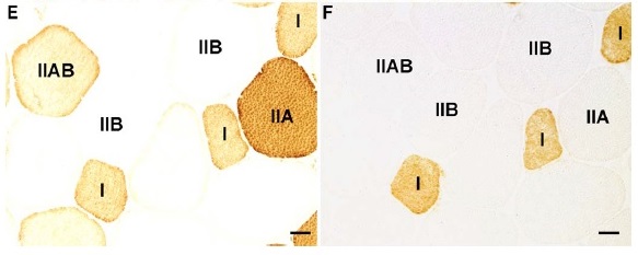
Interestingly, the subject who had the highest percentage of type IIB fibers (Fig. 1E, F), with approximately half of the muscle fibers being type IIB, had at the time for the biopsy the world record in his weight class in squat and he was not taking AS. Conversely, the subjects with the largest fiber areas were found in the PAS group, which means that these subjects in that respect had an advantage over those in the P group with smaller areas. Our study also shows that several subjects in the PAS group had a large amount of small fibers expressing fetal myosin. Adult muscle fibers do not normally express fetal myosin. The presence of developing myosin isoforms has been interpreted as signs of hyperplasia (McCormick and Schultz 1992; McCormick and Thomas 1992; Antonio and Gonyea 1993). A significant increase in fibers stained for developing isoforms of myosin, has been demonstrated with strength training (Antonio and Gonyea 1993; Kadi and Thornell 1999; McCall et al. 1996; Kadi 2000). These fibers might reflect newly formed fibers or abortively regenerated fibers. In the latter case, failed innervation can cause degeneration and the new fibers would be of no use for the athletes. It can be speculated that the increased number of fibers with fetal myosin is a result from using AS for almost ten years.
In conclusion, these results suggest that the action from AS is similar in the vastus lateralis, both long-term and 20 weeks supplemented, and in the trapezius muscles despite differences in contraction pattern. The results are in agreement according to larger fiber areas, correlation between myonuclei number per fiber and fiber area and to an increased number of myonuclei and satellite cells.
//juicedmuscle.com/jmblog/content/muscle-memory-fact-or-fiction
//www.juicedmuscle.com/jmblog/content/steroids-probably-result-lasting-benefit-its-users
- Login to post comments


